
Second generation cartilage cell implantation (3D chondrocyte implantation, matrix seeded autologous chondrocyte implantation ACI, MACI)
Second generation cartilage cell implantation is the latest technique for large focal chondral lesions. Cells are isolated from tissue explants and cultured in tissue engineering laboratory. Cells are seeded on a scaffold/matrix before reimplantation.
This procedure has many similarities with the first generation cartilage cell implantation, but it represents a newer version of it. The underlying principle of the method is that on site of the cartilage defects cartilage cells embedded in the extracellular matrix have only limited repair capacity. If cells are removed, extracellular matrix is digested, cells are isolated and cultured under regular tissue culture conditions cells demonstrate a good reproductive capacity and cell number will grow rapidly. Once the aimed cell number is reached cell suspension is not injected to the defect site, but cells are seeded on a special 3D matrix scaffold where cells would adhere to the fibers and start the production of the extracellular cartilage matrix components. As the cells grow, the material of the matrix decomposes and its space will be taken by the extracellular matrix.
Removal of the small cartilage specimens is done arthroscopically when a few rice body sized cartilage is collected in a sterile tube containing special transfer medium. Specimen is sent to the lab where immediate cell isolation and culturing is commenced. After 4-8 weeks cells would reach the appropriate number for cartilage repair and cells are seeded and attached to the scaffold with fibrin glue. Cells are cultured in this special 3D form for additional time.
A second surgery is performed with mini arthrotomy, the base of the cartilage defect is prepared, and the scaffold is cut to the appropriate size. These cartilage cell seeded scaffolds are secured with stitches and/or with fibrin glue providing appropriate environment (wetness) for the sample throughout the surgery.
The biggest advantage of the technique is that larger defect sizes (2-10cm2) may be repaired, and no severe donor site morbidity would occur.
Drawbacks of the technique is that two surgical intervention is necessary, the freshly developed cartilage is only hyaline-like tissue histologically although it appears to be superior when compared with the first generation chondrocyte implantation. This method is even more expensive than first generation ACI, the laboratory culturing is in the range of 6-8000€.
Postoperatively strict rehabilitation must be conducted by experienced physiotherapist, 6 weeks of partial weight bearing with crutches is followed by regular physiotherapy sessions. Full recovery should not be expected before 6 months postoperatively.
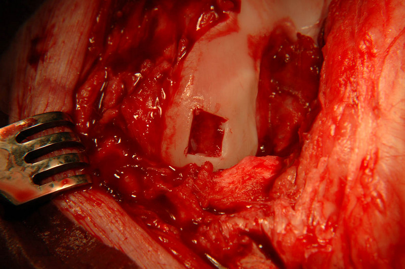 Cartilage defect of the femur
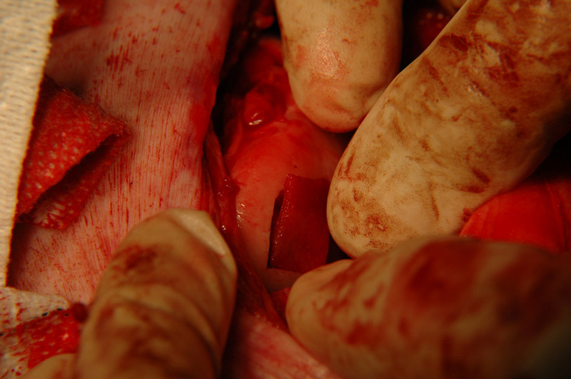 Placing the cell carrier matrix on the site of the cartilagedefect
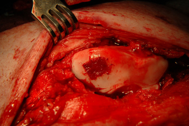 Matrix secured to the defect site using stitches and tissue glue